圳湾边贸易公司原装进口美国Gammex 156乳腺模体价格优惠!
厂家:Gammex, Inc.
品名:MammographicAccreditation Phantom Model 156
型号:Gammex 156, Gammex Model 156
------------------------
尺寸:
Dimensions
Phantom Body
Material . . . . . . . Acrylic
Phantom Dimensions . . . . . . . 4.5x10.2x10.8 cm (HWD) (1.75x4x4.25 in)
Acrylic Base . . . . . . . . . . . . . . 3.3 cm (1.3 in) thick
Cover . . . . . . . . . . . . . . . . . . . .0.3 cm (0.12 in) thick
Acrylic Disk . . . . . . . . . . . . . . . 4 mm thick x 1 cm diameter
Test Objects . . . . . . . . . . . . . . .Nylon fibrils (1.56, 1.12, 0.89, 0.75, 0.54 and
0.40 mm nylon fibers)
Simulated microcalcifications.(0.54, 0.40, 0.32, 0.24 and 0.16 mm specks)
Tumor-like masses . . . . . . . . .(2.00, l.00, 0.75, 0.50, 0.25 and 0.16 mm specks)
Included is a film, which is a contact image of the wax insert of your Gammex 156
----------------------
产品介绍:
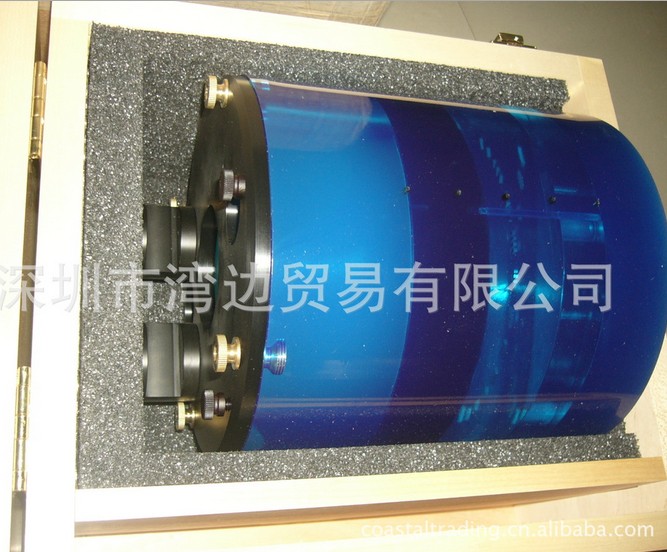
The Mammographic Accreditation Phantom Model 156 is
designed to test the performance of a mammographic
system by evaluation of the system’s ability to image
small structures similar to those found clinically. Objects
within the phantom were designed to simulate micro-calcifications,
fibrous structures in ducts, and tumor-like masses,
small structures that are important in the early detection
of breast cancer. Test objects within the phantom
range in size from those that should be visible on any system
to objects that will be difficult to see even on the best
mammographic systems.
Storage and Handling
Store the Mammographic Accreditation Phantom Model
156 in a cool dry area avoiding temperature extreme such
as intense heat and direct sunlight, or freezing temperatures.
Impact stress may cause breakage.
3
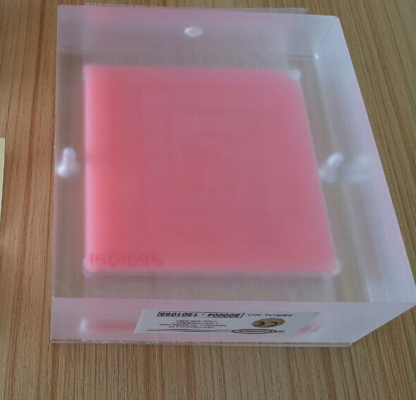
Product Description
The Mammographic Phantom is made up of a wax block
containing 16 various sets of test objects, a 3.3 cm (1.3
in.) thick acrylic base, a tray for placement of the wax
block, and a 0.3 cm (0.12 in.) thick cover. All of this
together approximates a 4.2 cm compressed breast. Five
groups of simulated micro-calcifications, six different size
nylon fibers simulate fibrous structures, and five different
size tumor-like masses are included in the wax insert.
Figure 1 lists the sizes of the test objects and their position
in relation to the notched corner of the wax block.
2 3 4
5 6 7 8
9 10 11 12
13 14 15 16
1
Figure 1: Location of the test objects in the wax insert.
Note: Numbers are for reference only.
4
Region Materials
1. 1.56 mm nylon fiber
2. 1.12 mm nylon fiber
3. 0.89 mm nylon fiber
4. 0.75 mm nylon fiber
5. 0.54 mm nylon fiber
6. 0.40 mm nylon fiber
7. 0.54 mm simulated micro-calcification
8. 0.40 mm simulated micro-calcification
9. 0.32 mm simulated micro-calcification
10. 0.24 mm simulated micro-calcification
11. 0.16 mm simulated micro-calcification
12. 2.00 mm thick tumor-like mass
13. 1.00 mm thick tumor-like mass
14. 0.75 mm thick tumor-like mass
15. 0.50 mm thick tumor-like mass
16. 0.25 mm thick tumor-like mass
5
A 4 mm thick, 10 mm diameter acrylic disk is also provided with
each phantom. The disk is used to establish and monitor a density
difference with respect to your facility’s operating level. It
should be placed on top of the phantom in a consistent location
in the image area so it does not obscure details in the phantom
and where it can not cast a shadow on any portion of the AEC
detector. According to the ACR Mammography QC Manual, a
suitable location is between and slightly below the first and second
largest fibers.
Using the Phantom*
Set Up
1. Center the phantom on the image receptor surface with the
nipple indent marker positioned away from the chest wall, just
as the nipple of the patient’s breast, and the chest wall edge
of the phantom aligned with the wall side of the image receptor.
2. Position the x-ray tube and compression device as you would
for a craniocaudal examination.
3. Select the mammographic machine settings appropriate for a
4.2 cm compressed breast. If the phototimer is used, set the
phototimer to the center position under the wax portion of the
phantom.
4. Make an exposure. Process the film. If using a digital mammographic
system, follow the manufacturer’s image processing
and display requirements.
Image Evaluation
The 156 Map conforms to the requirements of the American
College of Radiology for use in the ACR Accreditation Program.
The Phantom may be used to evaluate both screen/film and digital
images. ** Facilities participating in the ACR Accreditation
Program will use and evaluate the phantom images in accordance
with that program. Facilities following other national or
regional guidelines should refer to those standards.
1. Background optical should be greater than 1.40 O.D. for
screen/film images. The density difference between the
acrylic disk and the background should be at least 0.4.
Once the density difference is established as an operat
ing level, subsequent density difference measurements
should be within +/- 0.05 O.D.
2. The target mean glandular dose required to achieve the
optical density is as tabulated in the ACR Mammography
Quality Assurance manual for different anode/filter com
binations. Your alternative national or regional stan
dards.
3. Phantom images should be read under optimal condi
tions. Use of a X2 or greater magnifying glass or digital
image manipulation tools is recommended. Viewbox
luminance an ambient light levels should be optimized.
4. The ACR requires that the reader must be able to see all
of the 0.75 mm fiber, all of the 0.75 mm mass and all six
of the 0.32 mm specks. As many systems can surpass
this minimum, attention to degradation over time is as
important as meeting the minimum requirement.
Failure to meet the minimum image quality requirements set
either internally or by your accreditation or regulatory agency
should prompt investigation and corrective action.
You should consult with a medical physicist or engineer who
specializes in mammography and/or diagnostic radiology.
Following the correction, the system should be retested and the
new results documented.
*This manual provides a recommended use of the 156 phantom for routine
quality assurance in addition to, or in lieu of any other program.
**At the time of this writing, testing of the full field digital mammography systems
in the United States must follow the manufacturer’s quality control
manual.