|
|
|
|
INTRODUCTION
Spiral / Helical CT 2D Medium Contrast Phantom with four contrast stepsThe 2DMC provides the opportunity to evaluate low and medium contrast resolution in CT and particularly in Flatpanel-Detector-CT and Cone-Beam-CT (e.g. Dental-CBCT).
The Phantom has been designed to evaluate the imaging capabilities of 3D X-ray imaging modalities in the x/y-plane. CT-scanners low and medium contrast resolution capabilities can be obtained by a single scan using axial images and coronal reformations. The phantom visualizes the impact of all scan, image reconstruction, and display parameters. Specifications
3D Spatial Resolution PhantomThe D100-3DSR provides the opportunity to optimize collimation, pitch value and image reconstructio to achieve isotropic spatial resolution in all types of clinical applications.
The high-contrast spatial resolution test phantom visualizes the impact of collimation, slice width, pitch and image reconstruction algorithms. Spiral / Helical CT 3D Spatial Resolution PhantomMeasure in-plane and axial spatial resolution simultaneously. Optimize collimation, pitch value and image reconstruction to achieve isotropic spatial resolution in all types of clinical applications.
The high-contrast spatial resolution test phantom visualizes the impact of collimation, slice width, pitch and image reconstruction algorithms. Spiral / Helical CT 2D Low-Contrast Resolution Phantom with two contrast stepsMeasure the in-plane and axial low-contrast resolution. Optimize tube current, collimation, pitch and image reconstruction for the desired low-contrast resolution in all types of clinical applications.
The Phantom has been designed to evaluate the imaging capabilities of a CT scanner in the x/y-plane as well as in the axial-plane. The CT’s low-contrast resolution capabilities can be obtained by a single spiral scan using axial images and coronal reformations. The phantom visualizes the impact of all scan, image reconstruction, and display parameters. Specifications
Spiral / Helical CT Slice Sensitivity PhantomMeasure the sclice sensitivity profile (SSP) in spiral CT volume scans. Optimize collimation, pitch and image reconstruction for improved slice sensitivity and axial spatial resolution in all types of clinical applications.
Our phantom QRM-SSP is designed to test the slice sensitivity of a CT scanner’s spiral/helical scan modes. It contains a 25 micron thick metal foil of circular area (diameter 1 mm), embedded in a cylinder of uniform tissue-equivalent plastic. The heavy-metal insert is designed to evaluate all collimations from 0.5 mm to 10 mm with adequate image contrast. We suggest to analyze the maximum CT number of the high-contrast insert for a series of axial images. Specifications
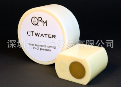
D100 Water TankQRM-Water-Tank provides the opportunity to evaluate noise and homogeneity in CT images. The water tank was designed to fit in the standard thorax and abdomen phantoms. It can be used as well to measure sample probes in water.
The Phantom can be attached to the standard QRM-Thorax and QRM-Abdomen, but can as well be used as a stand alone phantom. Specifications
Beam Stop PhantomBeam Stop Array for X-Ray Scatter Measurements.
The Phantom consists of a regular array of lead cylinders embedded in a polymethylmethacrylate (PMMA) plate. Under the assumption that the lead blockers offer an attenuation length sufficient to prevent primary radiation from reaching the detector, QRM-BeamStop is a convenient means to experimentally determine the x-ray scatter-to-primary ratio for a given measurement setup for analog and digital radiography. Specifications
|
|||||||||||||||||||||||||||||||||||||||||||||||||||||||||||||||||||||||||||||||||||||||||||||||||||||||||||||||||||
 |
|
|
||